Table of Contents
ToggleWhen it comes to skin bumps, not all are created equal. Enter the fibrous papule and the basal cell carcinoma—two skin conditions that can easily confuse even the most seasoned dermatology enthusiasts. While one might sound like a fancy appetizer at a gourmet restaurant, the other is a serious contender in the skin cancer ring.
Overview of Fibrous Papule and Basal Cell
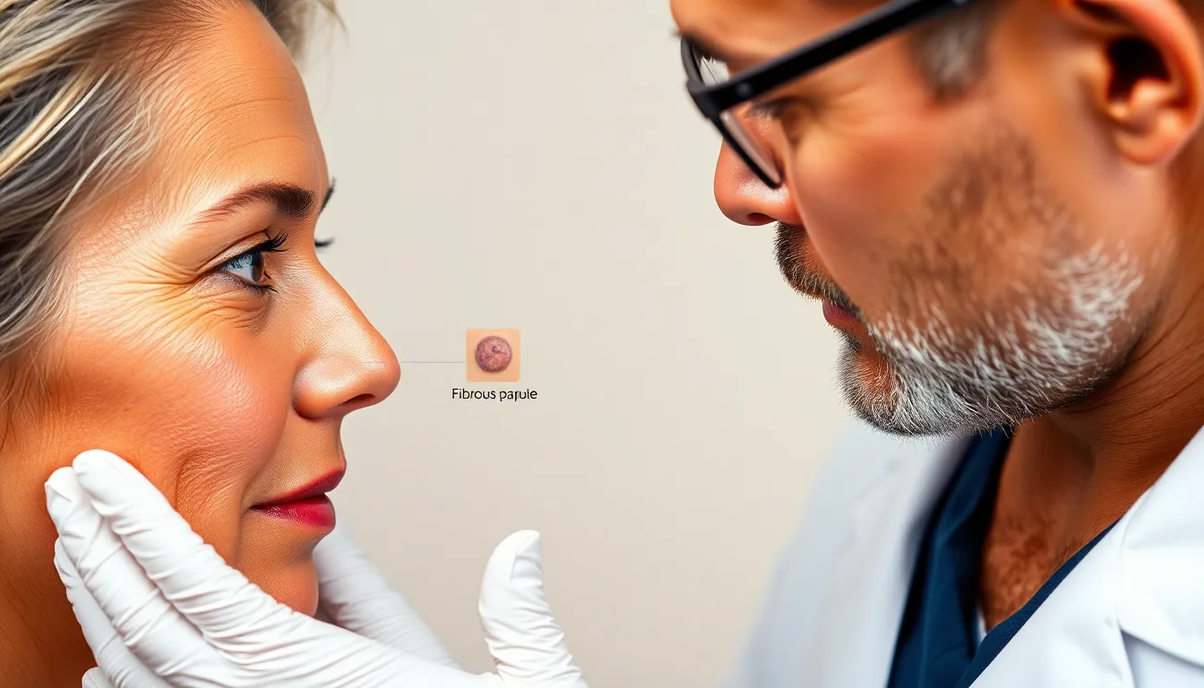
Fibrous papules appear as small, firm, flesh-colored nodules. Commonly found on the face, particularly around the nose, they usually occur in middle-aged adults. These benign tumors often do not require treatment unless for cosmetic reasons.
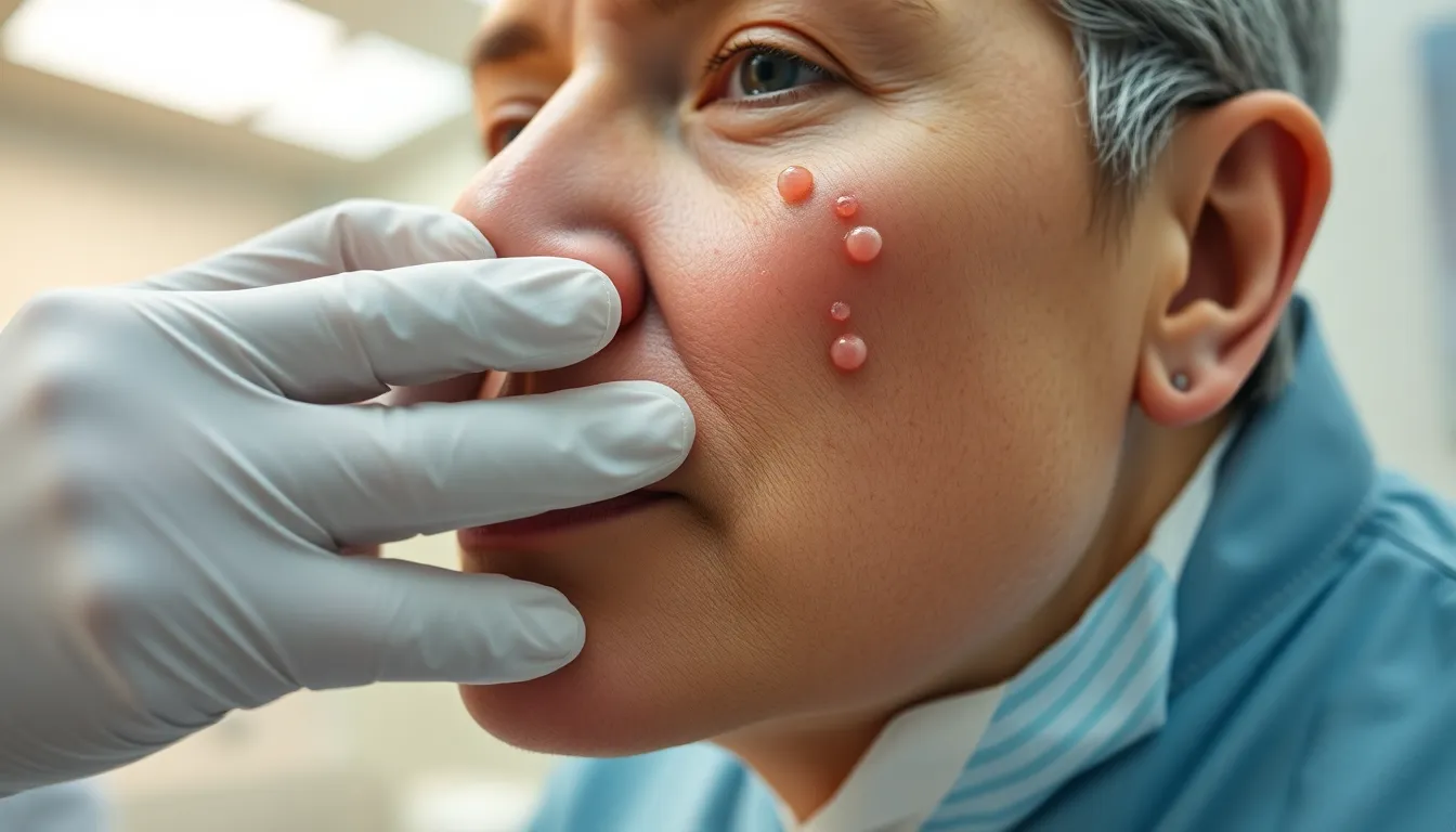
Basal cell carcinoma presents differently. It typically manifests as a pearly or waxy bump, often with a central depression. While basal cell carcinoma can occur anywhere on the body, sun-exposed areas are more susceptible.
Diagnosis usually involves a visual examination by a dermatologist. In cases of uncertainty, a biopsy may confirm the nature of the lesion. Recognition of the subtle differences is crucial for accurate assessment.
Fibrous papules generally pose no risk and are not associated with skin cancer. On the other hand, basal cell carcinoma carries a risk of local invasion. Patients might require surgical intervention for complete removal of malignant cells.
Understanding these conditions helps in making informed decisions regarding skin health. Recognizing the benign nature of fibrous papules can alleviate concerns for patients. Identifying basal cell carcinoma promptly may enhance treatment outcomes and reduce complications.
Knowledge about each condition’s characteristics aids in proper management strategies. They represent distinct variations of skin growths, requiring different approaches in terms of monitoring and treatment. Awareness fosters proactive skin care, promoting optimal dermatological health.
Differences Between Fibrous Papule and Basal Cell


Recognizing the differences between fibrous papules and basal cell carcinoma is essential for proper diagnosis and management.
Appearance and Characteristics
Fibrous papules appear as small, firm, flesh-colored nodules. These nodules typically measure 1 to 5 mm in diameter. Basal cell carcinoma presents differently, often resembling a pearly or waxy bump with a central depression. Bumps may exhibit telangiectasia, or tiny visible blood vessels, on their surface. While fibrous papules are benign and non-cancerous, basal cell carcinoma carries the risk of local invasion into surrounding tissues. An appropriate examination by a dermatologist can help differentiate these conditions effectively.
Common Locations
Fibrous papules frequently occur on the face, particularly on the nose and cheeks, common among middle-aged adults. In contrast, basal cell carcinoma tends to develop in sun-exposed areas. Locations such as the scalp, ears, neck, and back are often affected. The prevalence of sun exposure in these areas contributes to the higher likelihood of developing basal cell carcinoma. Identifying the common locations helps in recognizing and monitoring skin changes.
Causes and Risk Factors
Fibrous papules typically arise from genetic factors, often linked to familial tendencies. They commonly develop during adolescence and early adulthood, frequently appearing on the face. Skin types also play a role, where individuals with lighter skin exhibit a higher likelihood of developing such lesions. Age serves as another risk factor, with middle-aged adults showing more frequent occurrences.
Basal cell carcinoma’s etiology predominantly involves chronic sun exposure. UV radiation from the sun damages skin cells over time, leading to mutations. Areas exposed to sunlight, including the scalp, ears, neck, and back, show heightened incidence rates. Additionally, individuals with fair skin or those who burn easily face greater risks. Those with a history of frequent tanning bed use also have an increased likelihood of developing basal cell carcinoma.
Other risk factors include a personal or family history of skin cancer, which raises susceptibility levels. Patients with weakened immune systems due to medical conditions or medications are at higher risk as well. Exposure to certain chemicals, like arsenic and other toxic substances, may contribute to the development of basal cell carcinoma. Lastly, specific genetic conditions, such as Gorlin syndrome and albinism, significantly elevate risk profiles for both fibrous papules and basal cell carcinoma.
Understanding these causes and risk factors aids in recognizing the potential for each condition. A proactive approach to skin care, including regular check-ups and sun protection, helps reduce the chances of developing basal cell carcinoma.
Diagnosis and Identification
Diagnosis of fibrous papules and basal cell carcinoma relies on careful evaluation. A dermatologist typically conducts a thorough visual examination to differentiate between these conditions.
Clinical Examination
During a clinical examination, a dermatologist assesses the size, shape, color, and texture of the lesions. Fibrous papules appear as small, firm, flesh-colored nodules measuring 1 to 5 mm. In contrast, basal cell carcinoma manifests as a pearly or waxy bump, often featuring a central depression. Dermatologists also note the presence of telangiectasia, small visible blood vessels, on basal cell carcinoma. Observations regarding the lesion’s location provide additional context. Fibrous papules are primarily found on the face of middle-aged adults, while basal cell carcinoma frequently occurs on sun-exposed areas like the scalp, ears, neck, and back. Accurate clinical assessment allows healthcare professionals to make informed decisions regarding further diagnostic steps.
Biopsy and Histopathology
If the clinical picture is unclear, a biopsy confirms the diagnosis. Biopsies involve removing a small tissue sample from the lesion for microscopic examination. This process distinguishes fibrous papules from basal cell carcinoma based on histopathological features. Under the microscope, fibrous papules show fibrous tissue and collagen, which confirms their benign nature. Conversely, basal cell carcinoma reveals atypical keratinocytes and abnormal architecture, indicating potential malignancy. Accurate histopathological analysis provides essential information for treatment planning. Understanding these diagnostic methods enhances patient care and guides appropriate management.
Treatment Options
Effective treatment options for fibrous papules focus on cosmetic preferences. Many patients opt for removal due to aesthetic concerns rather than medical necessity. Common methods include cryotherapy, where liquid nitrogen freezes the lesion; electrosurgery, which uses electrical currents to remove the growth; and surgical excision for complete removal, especially if the lesion is larger or bothersome. These procedures generally result in minimal scarring.
In contrast, treatment for basal cell carcinoma is crucial due to its potential for local invasion. Surgical excision represents the primary method, ensuring complete removal of cancerous tissue. Mohs micrographic surgery provides a tissue-sparing option, allowing for precise excision while preserving healthy skin. Topical chemotherapy, such as imiquimod or 5-fluorouracil, offers an alternative for superficial lesions. Radiation therapy may apply for patients unable to undergo surgical procedures. Early intervention significantly improves patient outcomes and reduces recurrence risks.


